The cephalometry It remains a central tool in orthodontic diagnosis, even in an era dominated by CBCT, intraoral scanners, and digital planning.
The Steiner and Ricketts analyses are two of the most widely used systems for interpreting lateral skull radiographs, and they directly influence the treatment plan. orthodontics and dentofacial orthopedics.
Beyond simply "memorizing angles," understanding their differences, indications, and limitations is essential to avoid diagnostic errors that can lead to inappropriate treatment decisions (extractions, orthopedics, orthognathic surgery, etc.).
The importance of cephalometry in daily practice
The x-raycephalomethnic It allows the analysis of skeletal and dental relationships and, in certain analyses, also the soft tissue profile, using reproducible reference points and planes.
Its function is not to replace the clinical examination, but to objectify growth patterns, sagittal and vertical discrepancies, incisor position and inclination, and facial harmony, facilitating communication with the patient and between professionals.
In the practice of the general dentist who performs orthodontics, cephalometry helps to answer key questions:
- Is the problem primarily skeletal or dental?
- Is there a tendency towards anterior or posterior mandibular rotation?
- What margin do we have for dental compensation without compromising the facial profile?
These answers depend largely on how Steiner and Ricketts' analyses are interpreted.
Steiner's cephalometry: focus on sagittal and incisor relationships
Cecil C. Steiner He published his analysis in 1953 with a clear objective: to create a simple and clinical system, based on a few but very significant measures.
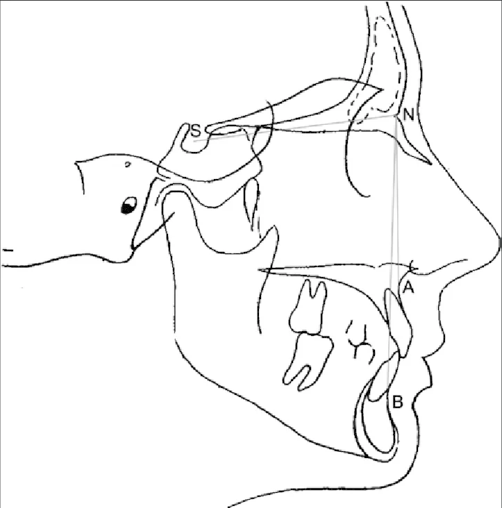
He replaced the Frankfort plan with the Sella plan–Nasion (SN) as a reference, due to its reproducibility and relative stability in growth, and structured the analysis into three blocks: skeletal (SNA, SNB, ANB), dental (incisor position) and, indirectly, profile osseous (Pg–NB).
Key skeletal measurements (SNA, SNB, ANB)
In Steiner, the SN plane serves as a basis for evaluating the sagittal position of the maxilla and mandible:
- SNA (82° ± 2°): anteroposterior position of the maxilla with respect to the cranial base.
- SNB (80° ± 2°): sagittal position of the mandible with respect to the same plane.
- ANB (2° ± 2°): SNA − SNB difference, used as a Class I, II or III skeletal classifier.
ANB values greater than 4° indicate a tendency towards skeletal Class II, while negative values suggest Class III, always considering the relative contribution of the maxilla and mandible.
Incisors and therapeutic commitment
Steiner places considerable emphasis on the position of the upper and lower incisors:
- U1–NA (angle and mm): proclination and protrusion of the upper incisor.
- L1–NB (angle and mm): proclination and protrusion of the lower incisor, fundamental in extraction decisions.
- Interincisal angle (~131° ± 6°): relationship between both incisors; low values indicate bimaxillary protrusion.
With these parameters, Steiner introduces the concept of "acceptable compromises": it is not always possible to take an ANB from 6° to 2°, but it is possible to adjust the position of the incisors to achieve a reasonable functional and aesthetic result on a non-ideal skeletal base.
Main limitations
Although very useful, Steiner's analysis has significant limitations:
- It depends on the SN plane, whose inclination can vary between patients and bias SNA, SNB and ANB.
- It does not offer a complete vertical assessment (hyper/hypodivergent pattern, facial height proportions).
- Its soft tissue analysis is limited; it needs to be complemented with other systems (Holdaway, Ricketts' E-line, etc.).
Ricketts cephalometry: growth, verticality and facial harmony
Robert M. Ricketts developed his analysis from thousands of cephalometric measurements processed using Rocky Mountain Data Systems, incorporating adjustments for age and a more comprehensive view of the patient.
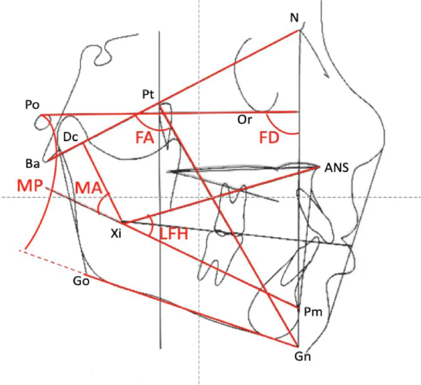
His proposal is based on the Ba–N (Basion–Nasion) plane and on concepts such as facial axis (PT–Gn), facial convexity and lower facial height, explicitly integrating growth patterns and biotype.
Facial axis and growth pattern
He facial axis (PT–Gn with respect to Ba–N) It is one of the pillars of Ricketts' analysis:
- Normatively it is situated around 90° ± 3.5°, and remains relatively stable with growth.
- Values >90° indicate anterior mandibular rotation (tendency towards a short face, deep overbite).
- Values <90° reflect posterior rotation (tendency towards a long face, open bite).
This stability of the facial axis makes Ricketts a very valuable tool for predicting growth and adjusting the long-term treatment plan.
Vertical dimension and facial proportions
Ricketts includes vertical measurements not considered by Steiner:
- Mandibular plane angle (FH–Go–Gn): discriminates hyperdivergent and hypodivergent patterns.
- Lower facial height (ANS–Xi–Pm): quantifies the proportion of the lower third of the face.
- Mandibular arch (Dr–Xi / Xi–Pm): linked to the rotation of the mandibular body.
These variables directly influence decisions such as vertical control with fixed appliances, the use of miniscrews, or the orthopedic/orthognathic approach.
Convexity and incisal position
The convexity (A to N–Pog) It is measured in millimeters and describes the sagittal profile:
- Rules regarding +2 mm ± 2 mm In childhood, with an approximate decrease of 0.2 mm/year due to anterior chin growth.
- Positive values indicate a tendency towards skeletal Class II; low or negative values indicate Class I/III with a flat or concave profile.
Ricketts also evaluates lower and upper incisors with respect to the A-Pog plane and FH:
- L1–A–Pog (mm and °) distinguishes bodily protrusion from simple proclination.
- U1–FH (111° ± 6°) It reflects the inclination of the upper incisor with respect to the cranial plane.
- The molar ratio (U6–PTV) It is adjusted to age, integrating the growth component into the sagittal assessment.
Steiner vs Ricketts: similarities, differences and complementarity
Both analyses share the basis of a lateral radiograph and standard bone points (N, A, B, Pog, ANS, Gn, Go, Me), but differ in their philosophy and clinical emphasis.
| Aspect | Steiner | Ricketts |
| Main reference plane | SN (Sella–Nasion), anterior cranial base. | Ba–N (Basion–Nasion) and FH, wider cranial base. |
| basic growth axis | It does not define a specific facial axis; it uses ANB and other angles. | PT-Gn facial axis versus Ba-N, stable and predictive. |
| Vertical dimension | Limited evaluation; requires further analysis for vertical pattern. | It includes lower facial height and mandibular plane angle. |
| Age adjustment | Essentially fixed rules. | Age-adjusted norms for convexity, mandibular plane, and molar position. |
| Incisors | U1–NA, L1–NB, interincisal angle; key in extraction decisions. | Incisors with respect to A-Pog and FH, differentiating body protrusion and proclination. |
| Soft profile | Chin prominence (Pg–NB); without complete soft tissue analysis. | It integrates facial profile and convexity measurements; it can be complemented with E-line. |
| Complexity / time | Quick, with few dimensions, ideal for chairside use. | More complex, with constructed points (Xi, PT, PTV) and multiple parameters. |
In clinical practice, it is common to use Steiner for rapid sagittal classification and dental decisions, complemented by Ricketts for vertical patterns, growth, and facial harmony.
Common errors in interpretation in Steiner
Knowing the most common sources of error helps to avoid incorrect diagnoses and inadequate treatment plans.
1. Relying solely on the ANB
The ANB is a derived value (SNA − SNB) and inherits the limitations of both angles:
- A high ANB may be due to a protruding maxilla, a receding mandible, or a combination of both; interpreting “Class II” without reviewing SNA and SNB individually is simplistic and dangerous.
- Changes in the inclination of the SN plane (more “flat” or “tilted” skull) can alter ANB without a true skeletal change.
2. Ignore the vertical component
Steiner does not directly assess facial height or the angle of the mandibular plane; relying on it as the sole analysis can lead to underdiagnosing hyperdivergent or hypodivergent patterns.
In a patient with normal ANB but a very high mandibular plane, the main problem will be vertical, not sagittal, and planning should be oriented towards controlling the vertical dimension rather than "correcting the Class".
3. Disconnect incisors from the side and airway
The tendency to "normalize" U1-NA and L1-NB without considering the impact on the soft tissue profile and the airway can result in poor aesthetic outcomes or undesirable effects on the pharyngeal space.
Cephalometry should be read in conjunction with clinical photographs and, when appropriate, airway studies (CBCT), not in isolation.
Common errors in interpretation in Ricketts
Ricketts's greater richness also opens the door to errors if his fundamentals are not respected.
1. Incorrect identification of complex points (PT, Xi, PTV)
Some points are not "purely" anatomical, but rather constructed from or dependent on structures that are difficult to visualize:
- PT in the pterygomaxillary fissure, Xi as the geometric center of the mandibular ramus and PTV as the constructed vertical plane.
- Poor location alters the facial axis, facial height, and molar position, distorting the overall reading.
2. Not applying age-appropriate rules
Convexity, facial height, and mandibular plane have norms that change with age; interpreting childhood values with adult standards generates overestimation of discrepancies or false classifications.[8][4]
For example, a convexity of +2 mm may be physiological at 9–10 years of age, while in adults it is usually closer to 0–1 mm.
3. Confusing proclination with body protrusion
Ricketts distinguishes between inclination (angle) and protrusion (distance) of lower incisors with respect to A–Pog:
- An incisor can be highly inclined but not very protrusive, or vice versa; the aesthetic and stability implications are different.
- Making extraction decisions based solely on angle, without analyzing body position, can result in profiles that are too flat or retruded.
Practical strategies to minimize interpretation errors
In the context of daily clinical practice, especially for general dentists who perform orthodontics, it is helpful to follow a series of systematic guidelines.
1. Standardize radiographic imaging
- Verify reproducible cephalometric position: Frankfort plane approximately horizontal, without exaggerated flexion or extension.
- Control magnification and always use the same equipment and protocol when performing comparative studies over time.
2. Check layouts and points before measuring
- Confirm the correct marking of key points: S, N, A, B, Pog, ANS, Gn, Go, Me, and in Ricketts, Ba, PT, Xi, Pm, U6.
- Correct any questionable layouts before calculating angles and distances; software does not correct marking errors.
3. Always read the measurements together.
- In Steiner: analyze SNA and SNB separately before interpreting ANB, and review incisor position in relation to the skeletal pattern.
- In Ricketts: integrate facial axis, mandibular plane, convexity and incisor position, always adjusting to the patient's age.
4. Complement the analysis and do not take any of them as absolute.
No analysis is complete on its own:
- In complex cases, combine Steiner and Ricketts with other systems (Bjork–Jarabak, Tweed, Wits) and with three-dimensional diagnosis (CBCT).
- Use clinical photography, intraoral scanning, and digital models to verify consistency between cephalometry and clinical reality.
Integrating classical cephalometry into the digital age
In contemporary practice, Steiner and Ricketts cephalometry does not compete with digital orthodontics, but is integrated as a common language to interpret and communicate complex cases.
Three-dimensional diagnosis with CBCT, systematic clinical photography, and 3D planning on intraoral scanners benefit from a solid cephalometric reading that helps prioritize goals and visualize the impact of dental and skeletal movements.
Training in orthodontics today involves learning to translate these ancl analysisbasics to the digital environment, maintaining diagnostic rigor while taking advantage of advanced planning tools.
Train in orthopedics and digital clinical orthodontics with university support
If you are interested in delving deeper into cephalometry (Steiner, Ricketts and other analyses), integrating radiographic diagnosis with CBCT and clinical photography, and mastering the complete digital workflow in orthodontics, it is important to choose a training program that combines solid theory, real clinical practice and current technology.

In this context, the Double European Master's Degree in Orthopedics and Digital Clinical Orthodontics (UCAM) from Koline Institute It is presented as a particularly interesting option for dentists who want to take a definitive step towards advanced orthodontics:
- Program blended learning, with online and face-to-face training in Malaga, designed to combine clinical practice and postgraduate studies.
- European double university degree, with academic backing and a total of 180 ECTS that consolidate your training path in orthodontics.
- Focus on three-dimensional diagnosis, CBCT analysis, clinical photography, dentofacial orthopedics, invisible orthodontics, and comprehensive digital treatment planning.
- Clinical practice from the early years, case tutoring and supplementary training in aligner systems, geared towards real practice as an orthodontist in Spain.
If you are a registered dentist and want to make the leap to a safer orthodontic practice, based on rigorous cephalometric diagnosis and digital tools, we invite you to learn more about the program and request information on places, requirements and schedule.
It is an opportunity to transform the way you diagnose and treat your orthodontic cases, with training designed for the real clinical practice that is carried out in Spain today.
Discover the main differences between specializing as an orthodontist in Spain through a university master's degree or private master's degree. Read now >